Ultrasound, also called ultrasound and ultrasound, is an imaging test that allows you to see any organ or tissue in your body in real time. When the test is done with Doppler, your doctor may observe blood flow in this area.
Ultrasound is a simple, quick and unrestricted procedure, can be done whenever your doctor deems it necessary and there is no need to wait between ultrasounds. However, it is important to check for recommendations for the scan, such as filling the bladder or taking medicines to remove excess gases, as this can make it difficult to visualize your organs.
- Ultrasound is an imaging exam that your doctor may indicate to identify changes in your organs.
- Therefore.
- This hotfix may be recommended for:.
Ultrasound should be performed in a laboratory, clinic or hospital, always under medical advice, to assist in the diagnosis or treatment of various situations. In addition, before taking the test it is necessary to know the preparation of the exam, since in some types of ultrasound, it may be necessary to drink a lot of water, fast or take medicines to eliminate gases, for example.
The ultrasound should be done with the patient lying on a stretcher, then a thin layer of gel should be placed on the skin and the transducer is placed on top of that gel, dragging the device over the skin. This device will generate images that are visible on a computer and should be analyzed by the doctor.
After completing the test, the doctor removes the gel with a paper towel and the person can go home. The test does not cause pain or discomfort, is easily accessible and is generally not an expensive test, being covered by several health plans, although it can also be performed by the SUS.
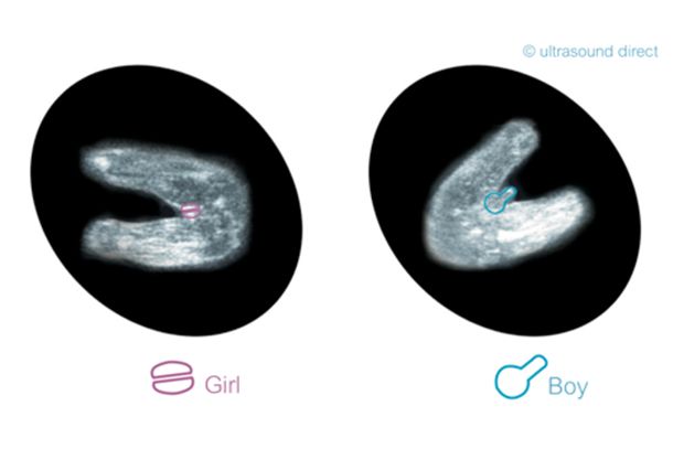
This is a particular type of ultrasound that should be done during pregnancy, between 20 and 24 weeks gestation, to check if the baby is developing properly or if he or she has any malformations, such as Down syndrome, myelomeningocele, anencephaly, hydrocephalus or congenital heart. Disease.
The duration of the test varies between 20 and 40 minutes and this test is recommended for all pregnant women.
How it’s done: Your doctor will place a gel in your pregnant woman’s belly and pass a device throughout the uterine region. The machine will generate images that can be viewed on the computer. Learn more about morphological ultrasound.
This is a type of examination that allows a better visualization of the structure to be studied, giving a more real look. 4D ultrasound, in addition to allowing excellent observation of the baby even in the maternal uterus, allows to capture their movements in real time.
They are especially suitable for viewing the fetus and can be taken from the 3rd month of pregnancy, but better images are obtained from the 6th month of pregnancy.
On breast ultrasound, your doctor may notice the appearance of a lump that may be felt when you feel your breast. This helps identify whether it may be a suspicious, benign mass or breast cancer, and is also useful for evaluating the breast ducts and looking for the causes of breast pain, for example.
How to do this: The woman should lie down without clothes or support while the doctor passes the equipment in all suspicious areas. It is normal to take longer to examine cysts or nodules. This test doesn’t replace mammography, but your doctor may prescribe it if the woman has large, firm breasts, making it difficult for mammography. Learn more about breast ultrasound.
On thyroid ultrasound, your doctor looks at the size of your gland, its shape, and whether you have nodules. This test may also be done to guide a biopsy so that a small sample of the tissue is taken, for example, in case of suspected cancer.
How to do this: The person should lie on his or her back, then place a gel on his neck. Your doctor will slide the device and you will see the person’s thyroid on the computer screen. It is normal for your doctor to ask if this is the first time the test has been performed or if there has been a change in previous tests to compare the results. Look for symptoms that may indicate thyroid cancer.
This test is indicated to visualize structures such as the uterus, ovaries, and blood vessels in this area, and may be needed to diagnose endometriosis, for example. It can be done by placing the transducer on the top of the belly or inside the vagina, in the latter case it is called transvaginal ultrasound. Learn the details of transvaginal ultrasound.
In men, a pelvic ultrasound is indicated to evaluate the prostate and bladder.
Abdominal ultrasound is used to study pain in the abdomen, if there are fluids in this area, or to assess organs such as the liver, kidneys, the presence of masses and in case of trauma or shock, in the stomach area. In addition to being useful when evaluating the kidneys and urinary tract for example.
How it is done: The doctor will indicate if any type of preparation is necessary before, but in the case of evaluation of the kidneys, urinary tract and bladder, before the test a 6-hour fast is recommended and the test should be done with the full bladder. Therefore, children aged 3 to 10 should drink 2 to 4 glasses of water, adolescents and adults should drink 5 to 10 glasses of water up to 1 hour before the test, unable to urinate before the test.