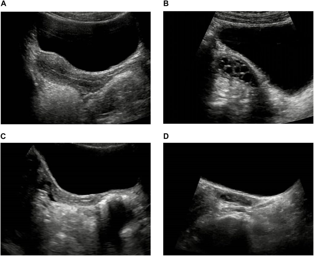
Morphological ultrasound, also known as morphological ultrasound or usG morphological ultrasound, is an imaging test that allows the baby to be seen inside the uterus, making it easier to identify certain diseases or malformations such as Down syndrome or congenital heart disease for example.
Ultrasound is usually indicated by the obstetrician in the second trimester, between week 18 and 24 of pregnancy, and therefore, in addition to fetal malformations, it may also be possible to identify the sex of the baby. In addition, the morphological USG marks the first time parents can see the developing baby in detail. Keep in mind that more tests should be done during the second trimester of pregnancy.
- Morphological ultrasound is used to identify the baby’s developmental phase.
- As well as to evaluate possible changes in developmental phases.
- In this way.
- The obstetrician can:.
In addition, when the baby is with his or her legs open, the doctor may also observe sex, which can then be confirmed by blood tests, for example. Refer to a list of available techniques to try to identify the sex of your baby.
A morphological ultrasound is recommended in the second trimester, between 18 and 24 weeks gestation, because it is when the baby is already sufficiently developed. However, this ultrasound can also be done in the first trimester, between week 11 and 14 of gestation, but because the baby is not yet well developed, the results may not be as satisfactory.
Morphological ultrasound can also be performed in the 3rd trimester, between 33 and 34 weeks gestation, but this usually only occurs when the pregnant woman has not undergone USG in the 1st or 2rd trimester, there is suspicion of malformation in the baby or when the pregnant woman has developed an infection that could interfere with the baby’s development. In addition to morphological ultrasound, 3D and 4D ultrasounds show details of the baby’s face and also identify diseases.
Morphological ultrasound in the second trimester can help identify several developmental problems in the baby such as spina bifida, anencephaly, hydrocephalus, diaphragmatic hernia, kidney disorders, Down syndrome, or heart disease.
See what your baby’s normal development should look like at 18 weeks
Normally, no special preparation is required to perform a morphological ultrasound, however, as the full bladder can help improve images and also raise the uterus, the obstetrician may advise drinking water before the test, as well as avoid emptying the bladder, if he or she feels like going to the bathroom.