Doppler ultrasound, also known as Doppler ultrasound or color eco-Doppler ultrasound, is an important test for evaluating blood vessel circulation and blood flow to a particular organ or region of the body.For example, your doctor may order it if a blood vessel is suspected to shrink, dilate, or occlude.
Some of the main indications of this test are evaluations of thrombosis, aneurysms or varicose veins, for example, and is also widely used during pregnancy, to check whether blood flow from the mother to the fetus occurs correctly, called fetal doppler.
- Like the common ultrasound exam.
- Doppler ultrasound is performed using a device capable of emitting sound waves.
- Which reach the tissue and return as an echo.
- Which is converted into images.
- Doppler is better able to identify and visualize blood flow at the site.
- Learn more about the main types of ultrasound and when they are indicated.
Doppler ultrasound is performed by your doctor at imaging clinics or hospitals and is available free of charge from the SUS or included in health plans.In particular, this review can cost between 200 and 500 reais, however, the price varies greatly depending on where it is performed.is done, the area observed or if there are complements to the exam, such as 3D technology, for example.
Some of the main situations in which a color doppler ultrasound is indicated are:
Sound waves generated during the exam produce the image directly on the computer screen of the device, so your doctor can see if there are any changes.
The Doppler ultrasound exam is simple and painless, it only requires lying on the stretcher while the doctor performs the exam.Fasting is generally not necessary, except for abdominal tests, such as artic doppler or kidney arteries.
In these cases, a 10-hour fast and the use of gas medications, such as dimethicone, may be indicated to reduce the formation of gases that may interfere with the test.
A color doppler ultrasound may be requested to evaluate virtually all areas of the body, however, some of the major doctor’s requests relate to:
Called lower limb doppler, it is often requested to identify varicose veins, thrombosis, narrowing of blood vessels, to evaluate blood flow before surgery in the area, or to assess the presence of symptoms of venous or arterial insufficiency, also known as poor circulation.
Understand what can cause poor circulation and major symptoms
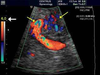
Also known as fetal Doppler, it is indicated by the obstetrician and is used to evaluate blood vessels and the rate of blood flow of the umbilical cord and placenta, observing whether there is a change in blood flow to the fetus, in order to plan better forms or delivery time.
This test is usually done in the third trimester of gestation, between 32 and 36 weeks, and is especially necessary if the doctor suspects alteration caused by situations such as low growth, maternal diabetes, changes in the amount of amniotic fluid, gestation of twins.or decreased fetal movements, for example.
Thyroid doppler may be indicated by the endocrinologist to evaluate the characteristics of thyroid blood vessels to help schedule punctures; It is also useful for identifying the malignancy characteristics of a nodule, as the presence of excess blood vessels may be another indication.of a suspicious nodule.
Learn more about when the thyroid nodule can be cancer
Carotids are arteries that carry blood from the heart to the brain, and when they undergo changes, such as obstruction or narrowing, they can cause symptoms such as dizziness, fainting or even a stroke.
Therefore, carotid doppler is indicated by the doctor in the face of suspicion of these changes, to assess the risk of stroke and also in people who have suffered a stroke, to help identify the cause. Find out what carotid ultrasound is for.
It is usually indicated by the nephrologist to study the flow of the renal arteries, seeking to identify the narrowings and occlusions of these vessels, which are causes of hard-to-control high blood pressure.
They may also be indicated to look for causes of kidney changes, such as decreased size, suspected aneurysms, or deformities.
It is indicated to assess the presence of dilations or aneurysms in the aorta, which may be suspected in people with abdominal breathing, it is also useful to study a dissection in this vessel, which is a serious complication caused by the detachment of its walls, or even observe the presence of atherosclerosis plaques that can cause a blockage of the aorta.
This test is also very important for planning correction surgery, if your doctor tells you to.Learn how to identify an aortic aneurysm and how to treat it.