Which is fetal echocardiography, how it is performed and when it is indicated
Fetal echocardiogram is an imaging exam usually requested during prenatal care and is designed to check fetal heart development, size, and function. Thus, it is able to identify certain congenital diseases, such as pulmonary atresia, atauricular or interventricular communication, as well as monitor the response to treatment in the case of arrhythmias for example. Find out what congenital heart disease is and its main types.
- This test does not require preparation.
- Is usually indicated from week 18 of gestation and is recommended for all pregnant women.
- Especially those over the age of 35 or who have a family history of congenital heart disease.
The test can cost between R$130 and R$400 depending on where it is done and whether it is done with doppler, however, the SUS makes it available and some health plans cover the review.
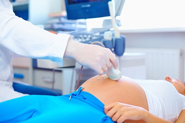
Fetal echocardiography is done in the same way as ultrasound, but only the baby’s heart structures, such as valves, arteries, and veins, are visualized, the gel is applied to the pregnant woman’s belly, which spreads with a device called a transducer, which emits waves that are treated, transformed into images, and analyzed by the doctor.
From the result of the scan, the doctor may indicate whether everything is fine with the baby’s cardiovascular system or indicate a heart disorder, thus determining whether treatment can be performed during pregnancy or whether the pregnant party should be referred to a hospital with an adequate structure to perform surgery on the fetus immediately after birth.
To complete the exam, no preparation is required and usually lasts about 30 minutes. It is a painless test that poses no risk to the mother or baby.
Fetal echocardiography is not recommended until week 18 of gestation, as the cardiovascular system and the visualization of the cardiovascular system are unclear by lack of maturation or even at the end of pregnancy, in addition the position, restlessness and multiple pregnancies make it difficult to examine.
Fetal doppler echocardiogram, in addition to allowing the visualization of fetal heart structures, also allows you to listen to the baby’s heartbeat, thus allowing to check if the heart rate is normal or if there is any indication of arrhythmia, which can be treated even during pregnancy. Understand what fetal Doppler is for and how it works.
The fetal echocardiogram should be done in conjunction with other prenatal tests and can be done from week 18 of gestation, during which it is already possible to hear the heartbeat due to the increased maturation of the cardiovascular system of the fetus. 18th week of pregnancy.
In addition to being indicated for prenatal care, this test is indicated for pregnant women who:
Fetal echocardiography is very important for all pregnant women because it helps identify heart changes in the baby that can be treated even during pregnancy immediately after birth, thus avoiding more serious complications.