Electrophoresis is a laboratory technique used to separate molecules according to their size and electrical charge in order to diagnose diseases, verify the expression of proteins or identify microorganisms.
Electrophoresis is a simple and economical procedure used in laboratory routines and research projects. Depending on the purpose of electrophoresis, more tests and tests may be needed to arrive at a diagnosis, for example.
- Electrophoresis can be performed for different purposes.
- Both in research projects and in diagnostics.
- As it is a simple and economical technique.
- Electrophoresis can therefore be performed to:.
Depending on the purpose of electrophoresis, more tests may be needed for your doctor to complete the diagnosis.
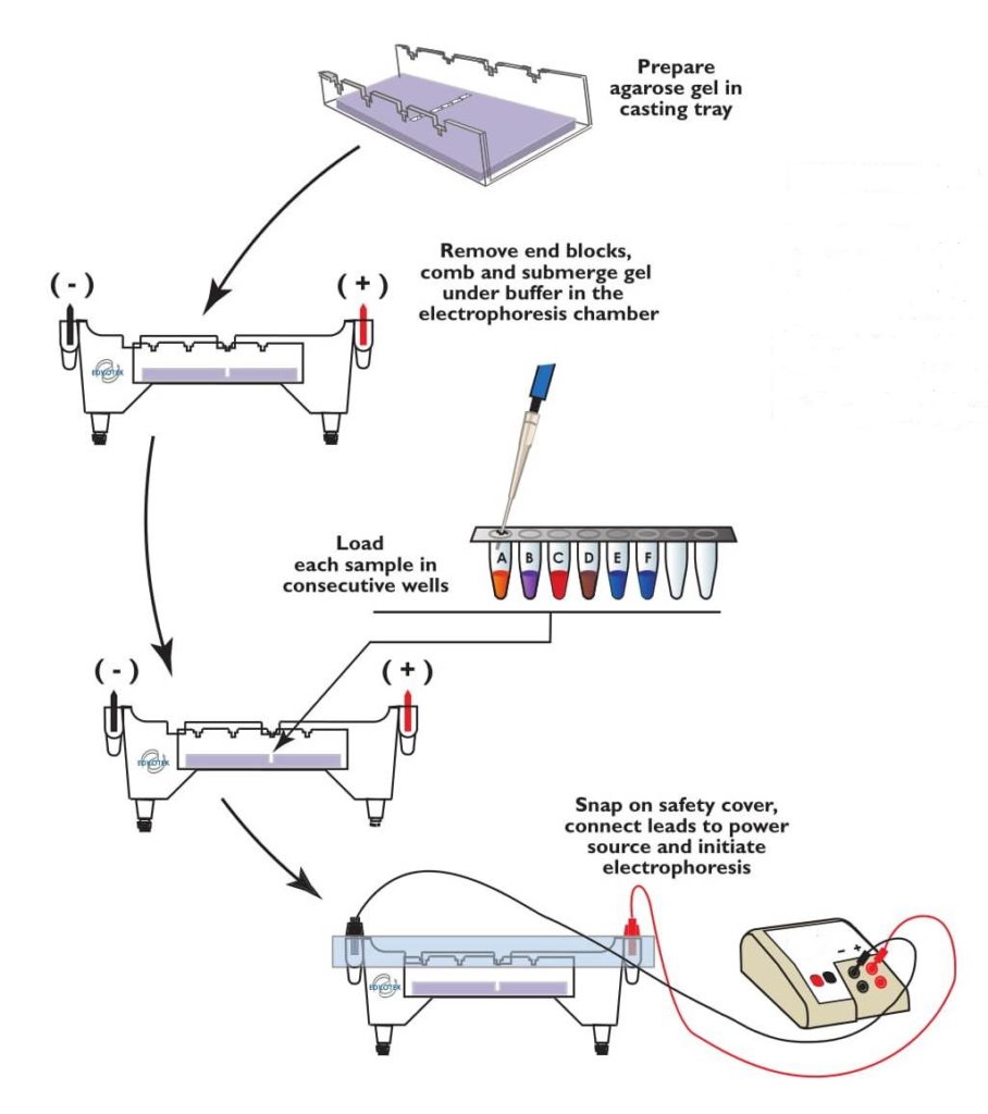
To perform electrophoresis it is necessary a gel, which can be polyacrylamide or agarous according to the lens, an electrophoresis pad and a tank, a molecular weight marker and a fluorescent dye, in addition to a UV or LED light equipment, also called a transiluminator. .
After preparing the gel, a specific object should be placed so that the wells are made into the gel, commonly called the comb, and let the gel take. When the gel is ready, simply apply the substances to the wells. To do this you must place a molecular weight marker in one of the wells, a positive control, which is the substance of which we know what it is, a negative control, which guarantees the validity of the reaction, and the samples to be analyzed. All samples must be mixed with a fluorescent dye, because in this way it is possible to visualize the strips in the transilluminator.
The gel with the samples should be placed in the electrophoresis tank, which contains the specific buffer solution, and then the device is turned on so that there is an electric current and therefore a potential difference, which is important for the separation of particles depending on its load and size. The electropholtic operating time varies depending on the purpose of the procedure and can last up to 1 hour.
After the specified time, it is possible to visualize the result of the electropholtic analysis through the translluminator. When the gel is placed under UV or LED light, it is possible to see the pattern of the strips: the larger the molecule, the lower its migration, approaching the well, while the lighter the molecule, the greater the migratory potential.
For the reaction to be validated it is necessary that the bands of the positive control are displayed and that the negative control does not show anything, because otherwise it is an indication that there has been contamination, and the whole process must be repeated.
Electrophoresis can be performed for different purposes and, depending on its purpose, several types of gel can be used, the most common being polyacrylamide and agarous.
Electrophoresis to identify microorganisms is more common in research laboratories, however, for diagnostic purposes, electrophoresis can be used to identify haematological diseases and diseases that evolve with increasing amounts of proteins, the main types of electrophoresis being:
Hemoglobin electrophoresis is a laboratory technique used to identify different types of hemoglobin circulating in the blood, identifying diseases related to hemoglobin synthesis. The type of hemoglobin is identified by electrophoresis at a specific pH, ideally between 8.0 and 9.0, checking a band pattern that can be compared to the normal pattern, allowing to identify the presence of abnormal hemoglobins.
Why it is made for: Hemoglobin electrophoresis is done to study and diagnose diseases related to hemoglobin synthesis, such as sickle cell disease and hemoglobin C disease, as well as being useful for differentiating thalassemia. Learn how to interpret hemoglobin electrophoresis.
Protein electrophoresis is a test requested by your doctor to assess the amount of protein circulating in your blood to identify diseases. This test is performed from a blood sample, which is centrifuged to obtain plasma, the part of the blood formed, among other substances, by proteins.
After electrophoresis, a band pattern and then a graph indicating the amount of each protein fraction can be visualized, being critical for diagnosis.
Why it’s made for: Protein electrophoresis allows your doctor to study the onset of multiple myeloma, dehydration, cirrhosis, inflammation, liver disease, pancreatitis, lupus and hypertension according to the band model and graph presented in the review report.
Understand how this is done and how to understand the outcome of protein electrophoresis.