3D or 4D ultrasounds can be performed during the prenatal period between week 26 and 29 and are used to view the physical details of the baby and assess the presence and severity of diseases, not only to reduce the curiosity of parents.
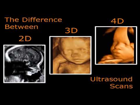
The 3D exam shows details of the baby’s body, allowing more clearly to see the face and genitals, while in the 4D examination, in addition to the well-defined features, it is also possible to visualize the movements of the fetus in the mother’s uterus.
- These tests can cost between R$200 and R$300.
- 00 and are performed in the same way as conventional ultrasound.
- Without the need for any special preparation.
- However.
- It is recommended not to use moisturizers in the stomach and drink plenty of fluids the day before.
- Proof.
The best time to do a 3D and 4D ultrasound is between weeks 26 and 29 of gestation, because during these weeks the baby has already grown and there is still a lot of amniotic fluid in the mother’s uterus.
Before this period, the fetus is still very small and low-fat under the skin, making it difficult to see its features, and after 30 weeks, the baby is very large and takes up a lot of space, making it difficult to see his face and movements.See also when the baby starts moving.
3D and 4D ultrasounds usually identify the same diseases as conventional ultrasounds and are therefore not normally covered by health plans. The main changes detected by ultrasound are:
The advantage of 3D or 4D testing is that they allow a better assessment of the severity of the problem, which can be done after diagnosis on conventional ultrasound.In addition, in most cases a morphological ultrasound is used, which is part of prenatal examinations that need to be done to identify diseases and malformations in the baby.Learn more about morphological ultrasound.
Some situations can interfere with images generated by 3D or 4D ultrasound, such as the baby’s position, which may be with the mother’s back, preventing the doctor from identifying her face, or the baby being with the limb or umbilical cord in front of the face.
In addition, the small amount of amniotic fluid or excess fat in the mother’s uterus can interfere with the image, in fact, excess fat makes it difficult to pass the waves that make up the image through the ultrasonic device, meaning that the formed images do not.reflect reality or don’t have a good resolution.
It is important to remember that the scan begins with a normal ultrasound, since 3D/4D ultrasound is only performed when good images are obtained during conventional scanning.