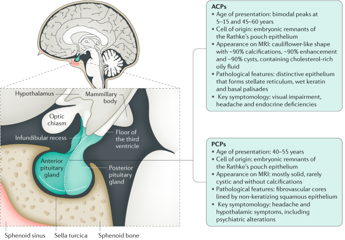
Craniopharynioma is a rare type of tumor, but it’s benign. This tumor affects the region of the Turkish chair, in the central nervous system (CNS), affecting a gland in the brain called the pituitary gland, which releases hormones to perform various functions of the body, and as the tumor grows, it can reach other parts of the body. and alter the functioning of the body.
There are two types of craniopharyniomas, adamantinomateux, which is the most common and affects more children than adults, and the papillary type, which is rarer and common in adults. Both come from a defect in brain cell formation, and symptoms are similar, with headaches, total or partial vision loss, growth problems in children and hormonal deregulation in adults.
- Treatment of this type of tumor may be done through surgery.
- Radiation therapy.
- Brachytherapy.
- And medication.
- Craniopharynyma has a difficult resection.
- But with proper treatment you can live with a better quality of life and with few neurological.
- Visual and endocrine sequelaes.
Although in some cases symptoms may appear suddenly, symptoms usually appear gradually. Some of them are:
In addition, craniopharynyma changes hormone levels, which can lead to irregular menstruation and difficulty maintaining or getting an erection and, in children, can cause stunting.
Because craniopharynioma is a rare type of tumor and causes symptoms similar to other diseases, it is often difficult to diagnose, and is discovered some time after the onset of symptoms. Therefore, as soon as symptoms appear, it is important to consult a neurologist, as an early diagnosis can lead to less aggressive treatment and reduce complications.
Diagnosis of a craniopharynioma is initially to evaluate symptoms and perform tests to evaluate vision, hearing, balance, coordination of body movements, reflexes, growth and development.
In addition, your doctor may recommend blood tests to test hormone levels, such as growth hormone (GH) and luteinizing hormone (LH), as changes in these hormones may be related to craniopharynyma. Learn more about luteinizing hormone role and test reference values.
To assess the exact location and size of the tumor, imaging tests such as MRI and CT scan are also shown. Although this is rare, in some cases, your doctor may recommend a biopsy to rule out cancer.
Depending on the size and location of the craniopharynyma, the neurologist and neurosurgeon will indicate the type of treatment, which may include:
In addition, research is underway, where new treatments and medications for craniopharynyma are being studied, and some hospitals and clinics are admitting people to try these treatments.
Treatment with hormone replacement medications should be carried out throughout life and, in addition, regular follow-up by an endocrinologist is also very important. In some cases, another surgery may be necessary, as the tumor may grow back.
Craniopharynioma, even after treatment, can cause changes in the body, as in most cases hormone levels remain altered, so it is very important to maintain the treatment recommended by your doctor. In addition, when it reaches a part of the brain called hypothalamus, it can cause severe obesity, developmental delay, behavioral changes, body temperature imbalances, excessive thirst, insomnia and increased blood pressure.
In addition, in more severe cases, when the craniopharynyma increases in size, it can cause blindness or clogging parts of the skull, resulting in fluid buildup and hydrocephalus. Learn more about hydrocephalus.
Craniopharynyma is incurable and therefore it is necessary to continue using drugs throughout life, due to hormonal complications, and undergo regular imaging and blood tests as recommended by the doctor, as the tumor may recur. Despite this, the treatments are becoming more advanced, allowing us to live longer and with a better quality of life.