Keratoscopy, also known as corneal topography or corneal topography, is an ophthalmological examination widely used in the diagnosis of keratoconus, which is a degenerative disease characterized by corneal deformity, which eventually takes on a cone form, with difficulty seeing and more sensitivity to light.
This examination is simple, is done in the office of the eye and consists of mapping the cornea, which is the transparent tissue in front of the eye, identifying any changes in this structure, the result of corneal topography can be indicated by the doctor immediately.after the test.
- Although more used in the diagnosis of keratoconus.
- Keratoscopy is also widely performed in the pre- and postoperative period of eye surgeries.
- Indicating whether the person is able to perform the procedure and whether the procedure had the expected result.
Corneal topography is performed to identify changes in the corneal surface, mainly to:
In addition, keratoscopy is a procedure widely performed in preoperative refractive surgeries, which are surgeries that aim to correct the alteration of the passage of light, but not all people with alterations in the cornea are capable of performing the procedure., As This is the case of people with keratoconus, due to the shape of the cornea, they cannot perform this type of surgery.
Therefore, in the case of keratoconus, the ophthalmologist may recommend the use of specific glasses and contact lenses and, depending on the degree of change in the cornea, may indicate the performing of other surgical procedures.Understand how keratoconas are treated.
Corneal topography can also be performed in the postoperative period, it is important to check if the alteration has been corrected and the cause of poor vision after refractive surgery.
Kerratoscopy is a simple procedure, performed in the ophthalmological office and lasting between 5 and 15 minutes, to perform this test it is not necessary to dilate the pupil, since it will not be evaluated, it may be recommended that the person does not wear contact lenses.contact lenses 2 to 7 days before the test, but this recommendation depends on the guidance of the doctor and the type of lens used.
To perform the examination, the person is placed in a device that reflects several concentric rings of light, called Placid rings, the cornea is the structure of the eye responsible for the input of light and, therefore, depending on the amount of light reflected, it is possible to check the curvature of the cornea and identify the changes.
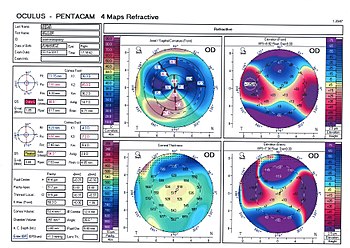
The distance between the reflected light rings is measured and analyzed by software on a computer associated with the computer, all information obtained from the emission of the rings of light is captured by the program and transformed into a color card, which must be interpreted by the doctor.Apart from the colors present, your doctor can check for changes:
Thus, the redder and orange the card, the greater the change in the cornea, indicating that more tests are needed to complete the diagnosis and begin appropriate treatment.