Calcific tendinitis occurs when there is a deposit of small calcium crystals in a tendon. This calcification can go away on its own, without treatment, but when used, ultrasound is used in physical therapy to remove calcium deposits, as surgery is necessary in most cases.
The cause of calcification should not be taken into account, but the theory is most affected by a dissolution of blood flow that inflames the inflamed tendon, inhabiting a calcium deposit there. Alterations in thyroid metabolism and estrogen can also promote their formation.
- It usually occurs from the age of 40 and is more common in women.
- Although it may appear on one side of the body.
- It could also affect both at once.
- One of the most affected tendons is the super spiny tendon.
- But the human rotator The cuff is also affected.
The only way to identify calcification in the tendons is through imaging tests. X-rays on the tendon wall, however, in case of calcification, you can see a small white area in the area from which it formed.
When the tendon is palpable, the person perceives a little pain, but it is not possible that there is only calcification for this reason, so an imaging test may be helpful, although it is usually not requested just for this suspicion.
Calcifying tendinitis often heals on its own, but there is spontaneous remission of bone deposit, however, it is not known when this will happen, so whenever a person develops symptoms he or she should perform treatment with physiotherapy sessions, using mucus instead of electrotherapy. to reduce inflammation and pain around you. Ultrasound is also able to reduce calcification with good results.
Painkillers and anti-inflammatory drugs contained in pills or ointments can also help fight pain, but in the most difficult cases, when no treatment relieves symptoms, if arthroscopy can be indicated, this surgery consists of a scraping of the calcified area, completely eliminating calcification. Infiltrations with anesthetics and corticosteroids are also indicated to relieve pain immediately, but this can only be done once or twice a year.
TenS and ultrasound are indicated for pain management, although it is not precisely separated from the latter, it acts on the reabsorption of deposited calcium, but if it increases the temperature of the area and blood flow, facilitating the elimination of calcium deposits.
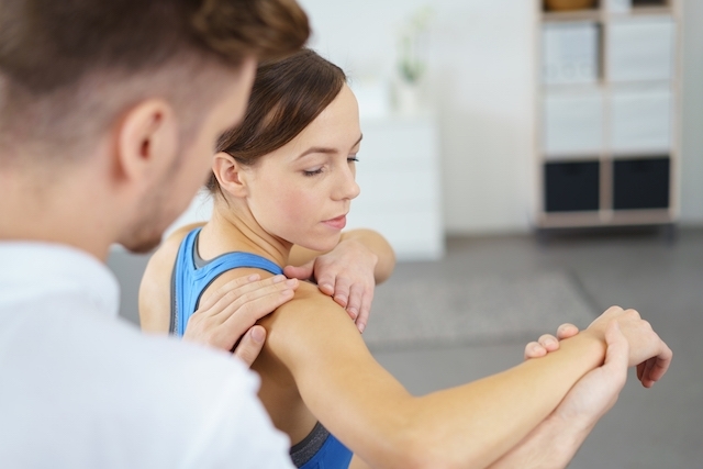
Trainings such as stretching and bodybuilding with elastic bands such as Thereband are indicated, as well as joint manipulation techniques. Pendulum exercises are very good strategies to reduce pain and maintain capsule integrity by avoiding a man’s protective position, resulting in more pain and a movement restriction.
The rest of the affected limb is indicated in case of pain and movement limitation, whenever it is possible to avoid carrying heavy objects with the affected arm, however, it is not necessary to have an absolute rest, so the use of a cable is not recommended, since it is important to maintain some movement to maintain the production of the synovial fluid that irrigates the joint.