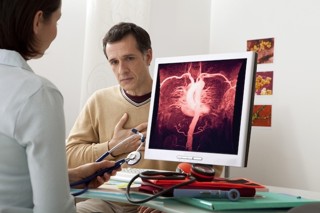
Angiotomography is a quick diagnostic test that allows the perfect visualization of fat or calcium plaques inside the walls and arteries of the body, using modern 3D equipment, very useful in coronary and brain diseases and that can also be invited to evaluate blood vessels in other parts of the body.
The doctor who normally requests this test from your cardiologist to evaluate the behavior of your heart’s blood vessels, especially if there are other cases of disturbance, such as heart spur, myocardial infusion scan, or for a heart pain assessment. For example.
- Angiotomography is used to clearly observe the inner and outer parts.
- Diameter and commitment of blood vessels.
- Clearly showing the presence of calcium or fat plaques in the coronary arteries.
- And also serves to visualize cerebral blood flow or any other part of the body.
- Such as the lungs or kidneys.
- For example.
This test can even detect minor coronary calcifications as a result of the buildup of fat plaques inside the arteries, which could not have been identified in other imaging tests.
The following table reflects some possible indications of different types of angiotomography:
To perform this test, a contrast is injected into the vase that is intended to be evaluated and the person must enter a CT team, which uses radiation to generate images that are displayed on the computer, so that the doctor can evaluate how the blood vessels, if there are calcified plates or if the blood flow is partially reduced.
Angiotomography lasts approximately 10 minutes and 4 hours before the individual has to eat or drink.
Daily medications can be taken at any time with water. It is recommended that you do not take anything that contains caffeine in any erectile dysfunction medication in the last 48 hours prior to the test.
A few minutes before angiotomography is done, some people may need to take medications to lower their heart rate, and others may need to dilate blood vessels; to improve the display of cardiac images.