The only way to confirm the diagnosis of glaucoma is to go to the ophthalmologist for tests to determine if the pressure inside the eye is high, which characterizes the disease.
Glaucoma tests are usually done when there are signs of suspected glaucoma, such as changes in routine eye examination, but they can also be prescribed as a prevention medium in people who are at increased risk of developing glaucoma, especially in cases of a family. history of the disease. .
Find out what the possible symptoms of glaucoma are and who is most at risk.
The main tests that an ophthalmologist may order to confirm the diagnosis of glaucoma include:
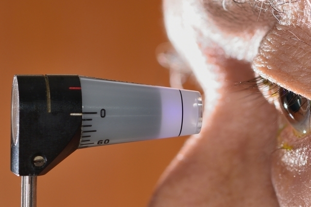
The eye pressure test, also called tonometry, assesses the pressure inside the eye, which in glaucoma is usually greater than 22 mmHg.
How it’s done: The ophthalmologist applies eye drops to numb the eye, then uses a device, called a tonometer, to apply light pressure to the eye to measure the pressure inside the eye.
The optic nerve evaluation test, scientifically called ophthalmoscopy, is a test that examines the shape and color of the optic nerve to identify if there are any injuries that may have been caused by glaucoma.
How it’s done: Your doctor applies eye drops to dilate your eye pupil, then uses a small flashlight to brighten your eye and look at your optic nerve, evaluating for any changes in your nerve.
The visual field assessment test, also called perimetry, helps the ophthalmologist identify whether there is a loss of the visual field caused by glaucoma, especially in the side view.
How is this done: In the case of the confrontational field, the ophthalmologist asks the patient to look straight ahead without moving the eyes and then passes a flashlight from side to side in front of the eyes, and the patient should warn each time that it is stop seeing the light. The most widely used, however, is automated perometry. See more details about the campimetry test.
The test used to assess the type of glaucoma is gonioscopy which determines the angle between the iris and the cornea, and when it is open it can be a sign of chronic open angle glaucoma and when it is narrow it can be a sign of angle glaucoma closed. either chronic or acute.
How it’s done: Your doctor applies anesthetic eye drops to your eye, then places a lens over your eye that contains a small mirror that allows you to observe the angle that forms between the iris and the cornea.
The cornea thickness assessment test, also called pachymetry, allows your doctor to understand whether the intraocular pressure reading, provided by tonometry, is correct or if you are affected by a very thick cornea, for example.
How to do it: The ophthalmologist places a small device in front of each eye that measures the thickness of the cornea.
Watch the video below and better understand what glaucoma is and the treatment options available:
In addition to the above tests, your eye doctor may also order more imaging tests to better evaluate eye structures. Some of these tests include: color retinography, Anteritra retinography, optical coherence tomography (OCT), GDx vcc, and HRT, for example.
If your glaucoma test has indicated that you have glaucoma, see how to treat glaucoma.
This test is used to guide you on your risk of developing glaucoma, based on your family history and other risk factors:
However, this test is not a substitute for a doctor’s diagnosis and consultation with an ophthalmologist is always recommended if glaucoma is suspected.