3D and 4D ultrasounds are imaging tests that can be performed during the prenatal visit between week 26 and 29 of gestation and are used to see the physical details of the baby and to assess the presence and severity of diseases, since they are only performed with the aim of reducing the curiosity of the priests.
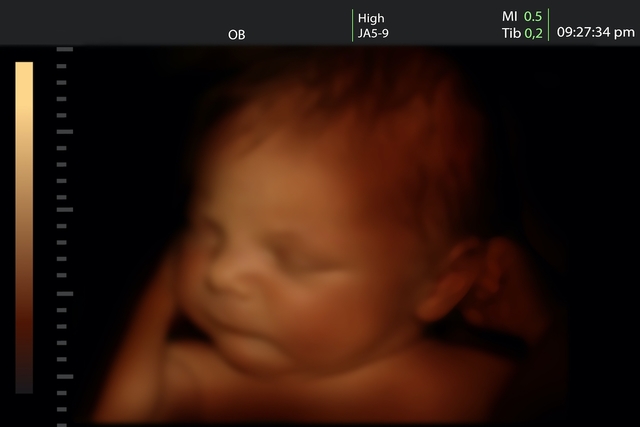
3D examination of the baby’s body details, although it is possible to see the face and genitals sharply, but during the 4D examination, in addition to the well-defined characteristics, it is also possible to visualize the movements of the fetus in the mother’s womb.
It is recommended not to use moisturizers in your stomach and drink plenty of fluids the day before the test.
The best time to do 3D and 4D ultrasounds is between weeks 26 and 29 gestation, but during those weeks when the baby grows, there is still amniotic fluid in the mother’s uterus.
Before this period, the fetus is still very small and has little fat under the skin, making it difficult to see its features, and after 30 weeks the baby is very large and takes up a lot of space, making it difficult to see her boyfriend and his movements. More details about how many months the baby begins to move in the mother’s womb.
Generally, 3D and 4D ultrasound identifies the same diseases as conventional ultrasound, so they are not normally covered by health plans. The main changes detected by ultrasound are:
The advantage of 3D 4D tests is that they allow a better assessment of the severity of the problem, which could be done after the diagnosis in conventional ultrasound, in addition, in most cases morphological ultrasound is used, which is part of the examinations prenatals. that must be gaps in the identification of diseases and malformations in babies. More information on structural or morphological ultrasound and how it should be performed.
Some situations can interfere with 3D or 4D ultrasound-generated images, such as the baby’s position, which could be returned by the mother, preventing the doctor from identifying her face, the fact that the baby may be with the umbilical cord on his or her face. .
In addition, the small amount of amniotic fluid, the excess fat in the mother’s face, could interfere with the image, this is because excess fat makes it difficult to form the waves that form the image in the ultrasound chamber, because images formed as a reflection of reality in the future have a good resolution.
It is important to remember that normal ultrasound scanning, because 3D/4D ultrasound is only performed when you get the best images from conventional scanning.